Human Anatomy and Physiology Laboratory Manual Review Sheet Exercise 1
Lab Exercise i: Introduction to Human Beefcake
- Anatomical Position
- Surface Beefcake
- Directional Terms
- Body Planes and Regions
Beefcake is the study of body structures. This can involve study of the large parts such equally muscle and organs similar the heart; called gross or macroscopic beefcake or, report of structures such every bit what heart muscle cells look similar with the aid of microscopes, microscopic anatomy. When we study what these structures do and how they do it, that is the realm of physiology. The prepare of exercises in Calendar week i is on the organization of the human body. What are the structures on the surface of the human body, how do we proper name them, how practice we describe their location(south) relative to each other and are we able to practice these with uniformity among anatomists and physiologists? These are some of the questions we aim to be able to reply by the end of the week's exercises.
Learning Outcomes
At the end of this lab, you should exist able to
- describe the anatomical position, and explicate its importance;
- use appropriate anatomical terminology to draw trunk regions, orientation and direction;
- recognize torso planes and be able to make up one's mind whether a department is in the frontal, transverse or sagittal aeroplane;
- proper noun the torso cavities, and indicate the important organs in each;
- name and describe the serous membranes of the ventral body cavities;
- identify the quadrants and nine regions of the abdomen on a torso model or image;
- list the 11 organ systems of the trunk;
- place major organs in the right organ system when presented with a list of organs.
Anatomical position
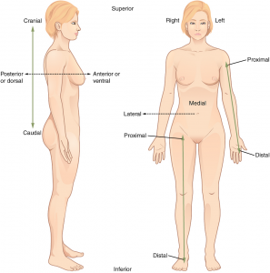
In clinical settings and when referring to specific areas of the human trunk, the body is oriented in a universal position called the anatomical position. In the anatomical position (Figure 1.1), the trunk is upright with the feet pointed forrard, eyes looking directly ahead, and artillery hanging at the sides with the palms facing forward. An individual in the anatomical position is said to be prone when lying face down and supine when lying face upward.
Directional Terminology
With the trunk in the anatomical position, specific terms are used to describe the location of a human body part relative to another (Figure 1.1a). A lateral view of an private in anatomical position (Figure 1. 1b) illustrates the front end and back orientation. Annotation that directional terms are with reference to the anatomical position.
(a) Lateral view (b) Inductive view
Figure one.1. Directional Terms Practical to the Homo Body Paired directional terms are shown as applied to the man torso. (Credit: CNX OpenStax CC BY four.0)
Click the link below to watch the an blitheness on anatomical position and directional terms:
https://www.wisc-online.com/learn/natural-science/life-science/ap15305/anatomical-terminology-relative-position
In quadrupeds (iv-legged animals), the directional terms are somewhat unlike.
| Tabular array 1.1. Directional Terms Used for Humans | ||
| Term | Pregnant | Example |
| Superior | Higher up | The mouth is superior to the chin |
| Inferior | Beneath | The umbilicus is junior to the breastbone |
| Cephalic/cranial | Toward the head | In humans, analogous to superior |
| Caudal | Toward the tail | In humans analogous to inferior |
| Anterior | To the front | The chest os is anterior to the lungs |
| Posterior | To the dorsum | The lungs are posterior to the breastbone |
| Medial | Toward the midline | The nose is medial to the cheek bones |
| Lateral | Abroad from the midline or medial plane | The kidneys are lateral to the navel |
| Ventral | Abdomen side | In humans, analogous to anterior |
| Dorsal | Back side | In humans, analogous to posterior |
| Superficial | Toward or at the body surface | The skin is superficial to skeleton |
| Deep | Abroad from the body surface | The dermis is deep to the epidermis |
| Proximal | Nearer the body or attached terminate | The elbow is proximal to the mitt |
| Distal | Farther from the trunk or attached end | The fingers are distal to the wrist |
Surface and regional anatomy
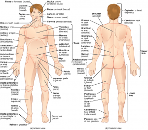
The torso is divided into ii primary regions, the axial (along the body's main axis) and appendicular regions (the appendages and structures attaching them to the main body centrality). Theaxial region includes the head, neck, and trunk; it runs forth the vertical axis of the body. Theappendicular region includes the limbs and the girdles that attach them to the trunk. These structures on the trunk surface constitute the body's surface beefcake; these are shown for the anterior structures (Effigy 1. 2a) and posterior structures (effigy i.2b) using the mutual and anatomical names. Body surface features are used every bit anatomical landmarks to assist in locating internal structures; as a result, many internal structures are named after an overlying surface structure.
Body Regions and Anatomical Landmarks
Figure 1.two. Regions of the Human Body The human torso is shown in anatomical position in an (a) inductive view and a (b) posterior view. The regions of the body are labeled in boldface. (Credit: CNX OpenStax CC Past four.0)
Click the link below to connect to the interactive object which enables learners to place a person's regional body parts.
https://world wide web.wisc-online.com/learn/general-education/anatomy-and-physiology1/ap17418/regional-body-parts
Body Planes and Sections
To obtain different views of the internal anatomy of an organ or trunk cavity; sections are made forth particular planes. At that place are Three primary planes along which such sections can be made; 2 of which are vertical and one horizontal (Effigy 1.3). The vertical planes are sagittal and frontal or coronal while the horizontal one is transverse. The sagittal cut or section divides the body into correct and left parts; when it passes through the midline of the body, information technology is called midsagittal because it divides into EQUAL right and left parts. A sagittal section that divides into Diff right and left parts is called parasagittal. The frontal cutting divides the trunk into anterior (front) and posterior (dorsum) parts and the transverse section divides the body into superior and inferior parts.
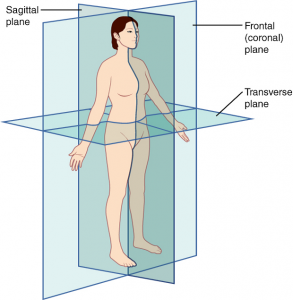
Figure 1.iiiPlanes of the Torso The three planes most ordinarily used in anatomical and medical imaging are the sagittal, frontal (or coronal), and transverse plane. (Credit: CNX OpenStax CC BY 4.0)
The body sections is narrated and illustrated in the animation; click on the link below:
https://www.wisc-online.com/learn/general-education/beefcake-and-physiology1/ap17618/body-sections-and-divisions-of-the-abdominal (CC-By-NC iv.0)
Major Body Cavities
Torso cavities are spaces within the body that surround internal organs. The torso cavities are covered with a serous membrane that supports and protects the organs it encloses. The serous membrane has two layers one nested to the organ chosen the visceral layer and the outer one called the parietal layer (Figure 1.4). The infinite betwixt the two layers chosen the serous cavity is filled with serous fluid which provides lubrication and reduces friction between the enclosed organ and side by side organs. The body cavities of a growing kid develop in the embryonic menstruum, during the fourth to eighth week of development.
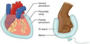
Figure i.ivSerous Membrane Serous membrane lines the pericardial cavity and reflects back to cover the heart—much the same manner that an underinflated airship would class ii layers surrounding a fist. (Credit: CNX OpenStax CC BY 4.0)
There are ii major torso cavities, dorsal and ventral. The dorsal cavity surrounds the encephalon and spinal cord.
The ventral body cavity is divided into the superior thoracic and inferior abdominopelvic crenel; each separated past the muscular diaphragm. The cavity is anterior to the vertebral cavalcade. The thoracic cavity contains the cavities surrounding the lungs, the left pleural cavity surrounding the left lung and correct pleural cavity surrounding the correct lung. The heart and the pericardial cavity surrounding information technology are located in an area betwixt the two pleural cavities called the mediastinum. The pericardial crenel lies in the anterior part of the mediastinum. The structures in the posterior and superior parts of the mediastinum include large blood vessels esophagus and trachea.
The thoracic crenel is separated from the abdominopelvic cavity by the diaphragm. The abdominopelvic cavity consists of the superior abdominal crenel that contains almost of the intestinal organs such equally the liver, gall float, pancreas, tummy, small intestine and large intestine. The junior pelvic cavity houses the urinary bladder internal reproductive organs parts of the large intestine including the rectum.
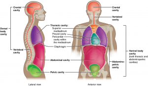
Figure 1.5 Dorsal and Ventral Body Cavities The ventral crenel includes the thoracic and abdominopelvic cavities and their subdivisions. The dorsal crenel includes the cranial and spinal cavities. (Credit: CNX OpenStax CC BY 4.0)
Click the link beneath to admission this interactive learning exercise. In this interactive object, learners examine the locations of major body cavities and their protective membranes. A drag-and-drib exercise completes the activity.
https://www.wisc-online.com/acquire/natural-science/life-science/ap15505/the-organization-of-the-human-body-torso-cavit
Abdominopelvic Quadrants and Regions
The abdominopelvic region can be further divided into iv quadrants or 9 regions to provide details about the internal abdominal and pelvic organs. The vertical and horizontal planes that separate the region to four quadrants bifurcate at the navel (umbilicus) while the two vertical and two horizontal planes that split up the region into straddle the navel. In clinical do the quadrants are used for reference while in anatomic studies the nine-region approach is often used. Identify or locate the quadrants and regions in Figure 1.6 and observe how they are named.
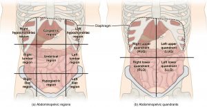
Figure 1.6Regions and Quadrants of the Peritoneal Cavity There are (a) 9 abdominal regions and (b) four abdominal quadrants in the peritoneal crenel. (Credit: CNX OpenStax CC BY four.0)
Organ Systems
We have seen the structures on the body surface and how they are named; and what organs or structures are situated in detail zones or regions of the body these constitute surface and regional anatomy respectively. In systemic anatomy, the organs making upward each of the ELEVEN organ systems and the functions of the systems are studied. Can you lot list the systems? The ii systems that influence the other ix to a marked caste are the nervous organization and endocrine system. The other systems include the largest organ system in terms of surface area covered, the integumentary system; muscular, skeletal, respiratory, cardiovascular, digestive, urinary, lymphatic and reproductive. The organ systems, some of the key organs and their functions are shown in Figure i.7.
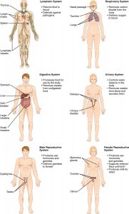
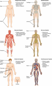
Figure 1.7Organ Systems of the Human being Body Organs that work together are grouped into organ systems. (Credit: CNX OpenStax CC BY 4.0)
sperlingforrawford.blogspot.com
Source: https://openlab.citytech.cuny.edu/sodeindebio2311/lab-1-human-body-organization/
0 Response to "Human Anatomy and Physiology Laboratory Manual Review Sheet Exercise 1"
Postar um comentário